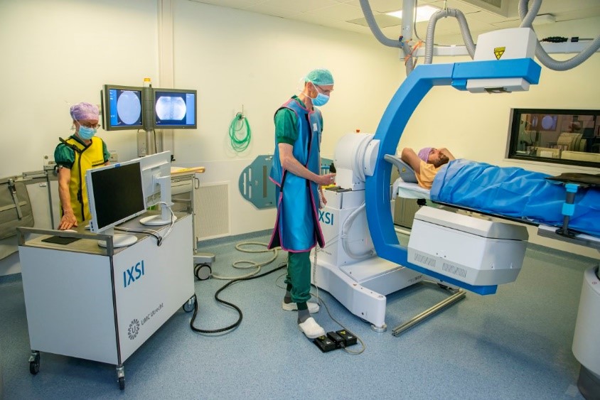
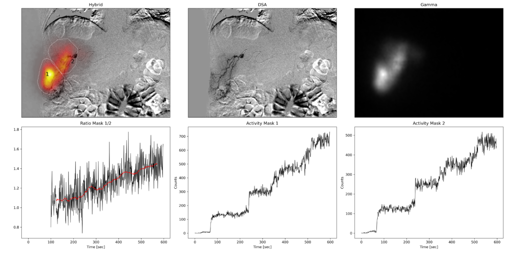
To demonstrate the clinical feasibility of hybrid imaging in the intervention room we have planned a clinical study involving the radioembolization pretreatment safety procedure using 99mTc-MAA. During a preparation or scout procedure MAA particles are injected in the liver. Next a diagnostic SPECT/CT at the nuclear medicine department is used to detect potential lung shunt of the particles, which may be a contraindication for the actual treatment. In addition the MAA distribution in the liver may be used to optimize the patients activity dose to be injected during the treatment procedure.
Objectives
The primary objective of the study is to demonstrate the safe application of hybrid imaging in the specific setting of the busy, dynamic intervention room. This will reveal potential, but unforeseen, risks to the patient and the medical personnel.
The secondary objective is i) to test the feasibility of IXSI to capture the dynamic process of injecting microspheres in a 2D movie for the first time ever and ii) to test the feasilbity of acquiring (quantitative) 3D SPECT/CT in the intervention room. This would also be unprecedented.
Study Design
The study foresees in including a maximum of 15 patients that are scheduled for clinical radioembolization of the liver. For this the mobile IXSI system will be introduced to a clinical x-ray intervention room or ‘angio suite’. After placing the catheter in the liver using the existing conventional x-ray C-arm, the interventional radiologist will switch to the IXSI modality that shows the same x-ray images but has nuclear capability as an extra. This will be used to record a movie of the injection of the MAA particles. After injection, a SPECT/CT of the liver and of the lungs is acquired. Then the patient switches back to the standard X-ray C-arm and the procedure is finished according to standard protocol. To manage potential risk or discomfort, the first 3 patients will receive no SPECT/CT yet.


